Neurological Disorders
•
46 likes•14,313 views

This document discusses spina bifida, a birth defect involving the neural tube where the spinal cord fails to close properly. It describes the three main types - spina bifida occulta, cystica (myelomeningocele), and meningocele. Signs and symptoms vary depending on location and severity of the defect but can include paralysis, loss of sensation, bowel/bladder issues, and cognitive problems. Treatment involves early surgery to repair the opening along with lifelong management of symptoms and secondary complications. Spinal cord injuries are also discussed, covering causes, classifications, regional effects, and long-term management strategies.
Report
Share


Neurological Disorders
- 1. Neurologic Disorders Ma. Tosca Cybil A. Torres, RN
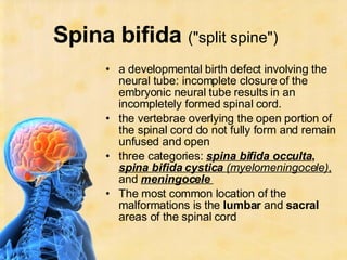
- 2. Spina bifida ("split spine") a developmental birth defect involving the neural tube: incomplete closure of the embryonic neural tube results in an incompletely formed spinal cord. the vertebrae overlying the open portion of the spinal cord do not fully form and remain unfused and open three categories: spina bifida occulta , spina bifida cystica (myelomeningocele) , and meningocele The most common location of the malformations is the lumbar and sacral areas of the spinal cord
- 3. Signs and symptoms varying degrees of paralysis absence of skin sensation poor or absent bowel and/or bladder control curvature of the spine (scoliosis) most cases there are cognitive problems Tethered Spinal Cord syndrome thought to result from scar tissue which forms following the initial surgery to close the open defect. Symptoms such as lower body pain, leg weakness, incontinence, scoliosis, numbness
- 4. types Spina bifida occulta Occulta is Latin for "hidden." one of the mildest forms of spina bifida although the degree of disability can vary depending upon the location of the lesion no opening of the back, but the outer part of some of the vertebrae are not completely closed split in the vertebrae is so small that the spinal cord does not protrude The skin at the site of the lesion may be normal, or it may have some hair growing from it; there may be a dimple in the skin, a lipoma, a dermal sinus or a birthmark
- 5. Spina bifida cystica ( myelomeningocele ) most serious and common form the unfused portion of the spinal column allows the spinal cord to protrude through an opening in the overlying vertebrae meningeal membranes that cover the spinal cord may or may not form a sac enclosing the spinal elements Superficially, the cyst may resemble an unrelated defect, sacrococcygeal teratoma the neural folds fail to meet and fuse leaving the spinal cord open and the involved area represented by a flattened, plate-like mass of nervous tissue with no overlying skin or membrane
- 6. The unfused elements of the spinal cord can be surgically closed along with the overlying muscle and skin shortly after birth there is usually some degree of paralysis and loss of sensation below the level of the spinal cord defect In approximately 90 percent of the people with myelomeningocele, hydrocephalus will also occur because the displaced cerebellum interferes with the normal flow of cerebrospinal fluid.
- 7. Meningocele least common form of spina bifida is a posterior meningocele (or meningeal cyst ) In a posterior meningocele, the outer faces of some vertebrae are open (unfused) and the meninges are damaged and pushed out through the opening, appearing as a sac or cyst which contains cerebrospinal fluid. The spinal cord and nerves are not involved and their function is normal. In an anterior meningocele , the inner faces of vertebrae are affected and the cyst protrudes into the retroperitoneum or the presacral space. causes of meningocele include teratoma and other tumors of the sacrococcyx and of the presacral space, and Currarino syndrome. Usually a meningocele has no negative long-term effects, although there are reports of tethered cord.
- 8.
- 9. Pathophysiology Spina bifida is caused by the failure of the neural tube to close during the first month of embryonic development (often before the mother knows she is pregnant) Causes: Medications such as some anticonvulsants, diabetes, having a relative with spina bifida, obesity, and an increased body temperature from fever or external sources such as hot tubs and electric blankets, unknown, genetic lack of folic acid (folate) is a contributing factor in the pathogenesis of neural tube defects
- 10. Medical-Surgical Treatment no cure for nerve damage to prevent further damage of the nervous tissue and to prevent infection, pediatric neurosurgeons operate to close the opening on the back During the operation, the spinal cord and its nerve roots are put back inside the spine and covered with meninges. In addition, a shunt may be surgically installed to provide a continuous drain for the cerebrospinal fluid produced in the brain, as happens with hydrocephalus. Shunts most commonly drain into the abdomen if spina bifida is detected during pregnancy, then open fetal surgery can be performed
- 11. Most affected individuals will require braces, crutches, walkers or wheelchairs to maximize their mobility. Many will need to manage their urinary system with a program of catheterization. They will need to insert a tube into their bladders to drain urine, of which the intervals vary from person to person, and may need medications to improve their dryness. Most will also require some sort of bowel management program.
- 12. Pregnancy screening Neural tube defects can usually be detected during pregnancy by testing the mother's blood (AFP screening) or a detailed fetal ultrasound. Spina bifida may be associated with other malformations as in dysmorphic syndromes, often resulting in spontaneous miscarriage. Genetic counseling and further genetic testing, such as amniocentesis, may be offered during the pregnancy as some neural tube defects are associated with genetic disorders such as trisomy 18. Ultrasound screening for spina bifida is partly responsible for the decline in new cases, because many pregnancies are terminated out of fear that a newborn might have a poor future quality of life. With modern medical care, the quality of life of patients has greatly improved.
- 13. Therapeutic management: Initial care: Monitoring of associated defects and complications Measures to prevent infection Monitoring of patency and functioning of VP shunt Adequate hydration and nutrition Normal infant stimulation Meticulous skin care Monitor I and O Emotional support to the family PT and ROM exercises; safelt measures for decreased sensation
- 14. b. Long term Orthopedic surgeons for treatment of contractures, gait analysis, and bracing Urologist for bladder and kidney function Pedia for minor infections, bowel program and coordination PT for maintenance of ROM OT for ADLs Intermittent catheterization for bladder control Weight maintenance diet to prevent obesity and maintain ambulation Counseling for long term adaptation Case management for long-term support of individual, family and school
- 15. Review of anatomy: Spinal Cord
- 16. Spinal cord injury causes myelopathy or damage to white matter or myelinated fiber tracts that carry sensation and motor signals to and from the brain. It also damages gray matter in the central part of the spinal, causing segmental losses of interneurons and motorneurons.
- 17. Causes: Trauma such as automobile accidents, falls, gunshots, diving accidents, war injuries, etc. Tumor such as meningiomas, ependymomas, astrocytomas, and metastatic cancer. Ischemia resulting from occlusion of spinal blood vessels, including dissecting aortic aneurysms, emboli, arteriosclerosis. Developmental disorders , such as spina bifida, meningomyolcoele, and other. Neurodegenerative diseases , such as Friedreich's ataxia, spinocerebellar ataxia, etc. Demyelinative diseases , such as Multiple Sclerosis. Transverse myelitis , resulting from spinal cord stroke, inflammation, or other causes. Vascular malformations , such as arteriovenous malformation (AVM), dural arteriovenous fistula (AVF), spinal hemangioma, cavernous angioma and aneurysm
- 18. Classification A indicates a "complete" spinal cord injury where no motor or sensory function is preserved in the sacral segments S4-S5. Since the S4-S5 segment is the lower segmental, absence of motor and sensory function indicates "complete" spinal cord injury. B indicates an "incomplete" spinal cord injury where sensory but not motor function is preserved below the neurological level and includes the sacral segments S4-S5. This is typically a transient phase and if the person recovers any motor function below the neurological level, that person essentially becomes a motor incomplete, i.e. ASIA C or D. C indicates an "incomplete" spinal cord injury where motor function is preserved below the neurological level and more than half of key muscles below the neurological level have a muscle grade of less than 3. D indicates an "incomplete" spinal cord injury where motor function is preserved below the neurological level and at least half of the key muscles below the neurological level have a muscle grade of 3 or more. E indicates "normal" where motor and sensory scores are normal. Note that it is possible to have spinal cord injury and neurological deficit with completely normal motor and sensory scores.
- 19. complications of spinal cord injury: Bowel and bladder function is regulated by the sacral region of the spine, so it is very common to experience dysfunction of the bowel and bladder, including infections of the bladder, and anal incontinence. Sexual function is also associated with the sacral region, and is often affected. Injuries of the C-1, C-2 will often result in a loss of breathing, necessitating mechanical ventilators or phrenic nerve pacing. Inability or reduced ability to regulate heart rate, blood pressure, sweating and hence body temperature. Spasticity (increased reflexes and stiffness of the limbs). Neuropathic pain. Autonomic dysreflexia or abnormal increases in blood pressure, sweating, and other autonomic responses to pain or sensory disturbances. Atrophy of muscle. Superior Mesenteric Artery Syndrome Osteoporosis (loss of calcium) and bone degeneration. Gallbladder and renal stones.
- 20. Cervical injuries Cervical (neck) injuries usually result in full or partial tetraplegia ( Quadraplegia ). C3 vertebrae and above : Typically lose diaphragm function and require a ventilator to breathe. C4 : May have some use of biceps and shoulders, but weaker C5 : May retain the use of shoulders and biceps, but not of the wrists or hands. C6 : Generally retain some wrist control, but no hand function. C7 and T1 : Can usually straighten their arms but still may have dexterity problems with the hand and fingers. C7 is generally the level for functional independence.
- 21. Thoracic injuries Injuries at the thoracic level and below result in paraplegia . The hands, arms, head, and breathing are usually not affected. T1 to T8 : Most often have control of the hands, but lack control of the abdominal muscles so control of the trunk is difficult or impossible. Effects are less severe the lower the injury. T9 to T12 : Allows good trunk and abdominal muscle control, and sitting balance is very good.
- 22. Lumbar and Sacral injuries The effect of injuries to the lumbar or sacral region of the spinal canal are decreased control of the legs and hips, urinary system, and anus.
- 23. Central Cord and Other Syndromes Central cord syndrome is a form of incomplete spinal cord injury characterized by impairment in the arms and hands and, to a lesser extent, in the legs. This is also referred to as inverse paraplegia , because the hands and arms are paralyzed while the legs and lower extremities work correctly. Most often the damage is to the cervical or upper thoracic regions of the spinal cord, and characterized by weakness in the arms with relative sparing of the legs with variable sensory loss. This condition is associated with ischemia, hemorrhage, or necrosis involving the central portions of the spinal cord (the large nerve fibers that carry information directly from the cerebral cortex). Corticospinal fibers destined for the legs are spared due to their more external location in the spinal cord. This clinical pattern may emerge during recovery from spinal shock due to prolonged swelling around or near the vertebrae, causing pressures on the cord. The symptoms may be transient or permanent.
- 24. Anterior cord syndrome an incomplete spinal cord injury. Below the injury, motor function, pain sensation, and temperature sensation is lost; touch, proprioception (sense of position in space), and vibration sense remain intact. Posterior cord syndrome can also occur, but is very rare.
- 25. Brown-Séquard Syndrome usually occurs when the spinal cord is hemisectioned or injured on the lateral side. On the ipsilateral side of the injury (same side), there is a loss of motor function, vibration, and light touch. Contralaterally (opposite side of injury), there is a loss of pain, temperature, and deep touch sensations.
- 26. Diagnostic: Clinical evaluation: absence of reflexes, flaccidity, loss of sensation below injury level Spinal x-ray: vertebral fractures, bony overgrowth CT scans/MRI: evidence of cord compression and edema or tumor formation
- 27. Therapeutic management: Surgery- laminectomy or fusion for decompression and stabilization, wound debridement, placement of cervical tongs or halo traction for stabilization, tracheotomy for mechanical ventilation as needed medications: massice corticosteroid therapy to improve outcome, vasopressors for shock, prophylactic antibiotics for open wounds, analgesics for pain, anticoagulants to prevent emboli and thrombus formation, anti anxiety to reduce emotional stress.
- 28. c. General: a. initial: 1. spinal stabilization with backboard or cervical collar on initial transport 2. MV if necessary 3. monitor cardiac status, blood gases, neuro V/S, I&O, V/S 4. maintain skeletal traction and body alignment 5. reposition and turn every 2hrs 6. passive ROM 7. monitor bowel and bladder function, skin integrity and avoid extreme temperatures
- 29. b. Long term 1. bowel training 2. bladder training 3. PT to diminish orthostatic hypotension, increase strength and endurance, decrease muscle spasticity, prevent contractures 4. OT to aid adaptation of ADLs 5. respiratory therapy 6. recreational therapy 7. speech therapy 8. case mgt for needed resources 9. long term medical ff up 10. counseling of individual and family support adaptation
- 30. Prevention and promotion: Daily skin inspections Diligent use of bowel and bladder programs to prevent bowel obstruction and UTI Influenza and pneumonia vaccines to prevent respiratory complications Early recogniton and treatment of urinary tract and respiratory problems