This article covers
What is the endoplasmic reticulum (ER)? A quick overview
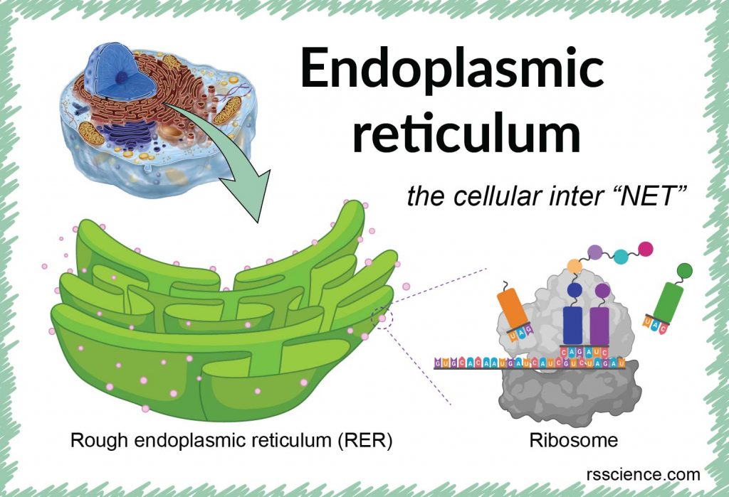
The endoplasmic reticulum (ER) is thought to be an internal membrane that divides into a branching web throughout the cytoplasm. The ER occurs in two morphologically distinct forms: rough ER and smooth ER.
The outer side (facing the cytosol) of the rough ER is studded with ribosomes that are the sites of protein synthesis. Under the electron microscope, the dense granular ribosomes gave the name “rough” ER. On the other hand, the smooth ER lacks ribosomes.
ER plays many important roles in our cells. In brief, rough ER coordinates protein synthesis and transportation. Smooth ER specializes in lipid synthesis, steroid hormone production, and detoxification. The lumen of ER is also the important reservoir of calcium ions (Ca2+) in the cells.
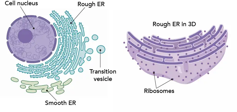
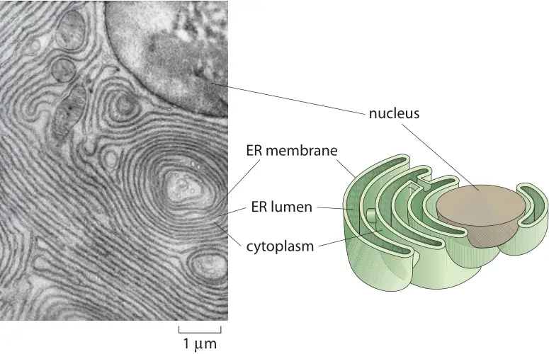
[In this figure] The anatomy of ER.
Left: The relationship between the nucleus, rough, and smooth ER. The newly synthesized proteins will be transported to the Golgi apparatus in transition vesicles. Right: A 3D view of rough ER. You can see the ER membrane actually forms branching networks of many interconnected sacs and tubes.
The image was created with BioRender.com.
The structure of endoplasmic reticulum
The endoplasmic reticulum (ER) is a large, dynamic interconnected network inside the eukaryotic cells. ER can be morphologically divided into two structures: rough endoplasmic reticulum (RER), and smooth endoplasmic reticulum (SER).
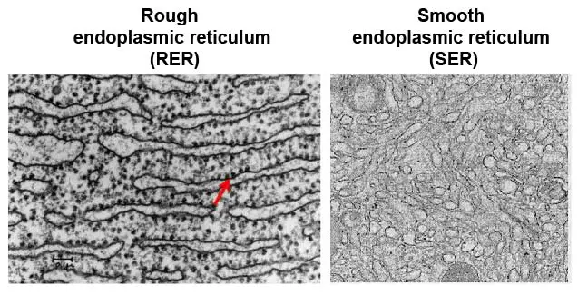
[In this figure] Left: Electron micrograph (EM) of rough ER. Red arrow indicates a ribosome particle on the cytoplasmic surface of rough ER. Right: EM of smooth ER.
Photo source: https://www.ncbi.nlm.nih.gov/pmc/articles/PMC4700099/
The surface of the rough endoplasmic reticulum (RER) is studded with protein-manufacturing ribosomes giving it a “rough” appearance (hence its name). The ribosomes attach to RER outer (cytoplasmic) surface. RER consists of an interconnected network of flattened, membrane-enclosed sacs known as cisternae or ER sheets. Cisternae are usually observed in a stacked configuration and are connected. The luminal spacing is very consistent, usually about 50 nm in mammal cells. Rough ER lies immediately adjacent to the cell nucleus, and its membrane is continuous with the outer membrane of the nuclear envelope.
The smooth endoplasmic reticulum (SER) consists of tubular structures, which are located near the cell periphery. SER has no ribosome on its surface, resulting in a smooth surface.
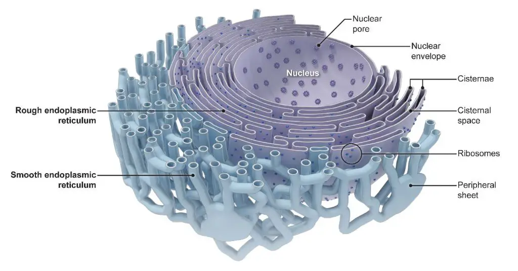
[In this figure] The structure and relationship of the nuclear envelope, rough, and smooth endoplasmic reticulum.
Rough ER almost always appears as parallel stacks of double membranes (known as cisternae, which are long, flat, and unbranched) that are heavily dotted with ribosomes. On the other hand, the smooth, tubular network (irregularly branched) characterizes smooth ER. The ER is attached to the double-layered nuclear envelope and provides a connection, or bridge, between the nucleus and the cytosol.
Photo source: https://elifesciences.org/articles/20468
The ER network is dynamic, continually rearranging, and growing. The ratio of sheets (RER) to tubules (SER) varies in different cell types and reflects the different functions of these cells. For example, the ER architecture of specialized cells that produce vast amounts of secreted proteins, such as pancreatic secretory cells and B cells, is largely made up of RER.
In contrast, cells that are involved in lipid synthesis and calcium storage possess an ER composed of primarily tubules. Adrenal, liver, and muscle cells are all examples of specialized cells with a predominantly tubular network and reflect the function of these cells. The endoplasmic reticulum is not found in red blood cells or sperm cells.
Endoplasmic reticulum membranes
The membranes of the endoplasmic reticulum are very similar to the cell membrane (also known as the plasma membrane). The backbone structure of these membranes is a thin polar membrane made of two layers of lipid molecules, called lipid bilayer (or phospholipid bilayer). This bilayer is formed by the molecular nature of amphiphilic phospholipids.
Phospholipids have a hydrophilic (preferring water) phosphate head and a hydrophobic (preferring to stay away from water) tail consisting of two fatty acid chains. In an aqua environment, the hydrophobic tails of many phospholipids naturally stay together with their hydrophilic phosphate heads facing the outside water molecules. The lipid bilayer forms spontaneously by self-assembly.
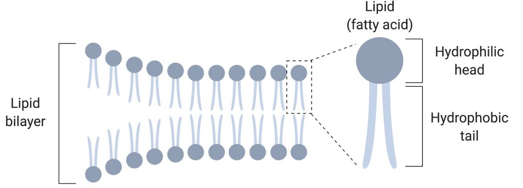
[In this figure] Amphiphilic phospholipids spontaneously form two lipid films, called lipid bilayer.
Lipid bilayer membranes naturally possess the property of selective permeability. While uncharged molecules and gases can easily cross the cell membrane, ions, proteins, and other charged molecules are impermeable. Therefore, lipid bilayer membranes can form compartments that keep distinct environments separated by the membrane barriers.
Since the lipid bilayers are so great to create compartments for biochemical reactions, the membrane-bound organelles (such as the nucleus, endoplasmic reticulum, mitochondria, chloroplasts, Golgi apparatus, lysosomes, peroxisomes, and vacuoles) all use the same lipid bilayers as their membranes.
Like cell membrane, there are many membrane-associated proteins on the ER membranes. They play several key functions like anchoring ribosomes on RER, transporting ions and proteins between ER lumen and cytoplasm, and maintaining or arranging the shape of ER structures.
What does endoplasmic reticulum do? – endoplasmic reticulum function
The endoplasmic reticulum is literally the place where the cellular factories locate. RER with the ribosomes is mainly involved in protein synthesis while the SER is mainly involved in lipid and steroid synthesis.
The function of rough ER
A ribosome is a particle-like cell organelle that serves as the site for protein synthesis in the cell. The ribosome reads the sequence of the messenger RNA (mRNA) and, using the genetic code, translates the sequence of RNA into a sequence of amino acids (a process called Translation).
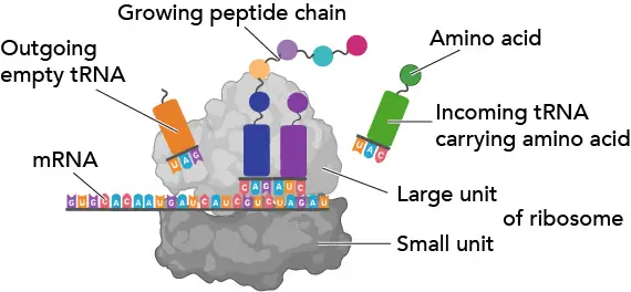
[In this figure] An analogy for ribosomes in a factory.
Ribosomes work like machines to translate the code sequence of mRNA into a protein. Scientists call ribosomes, the molecular micro-machines, to admire how exquisite the ribosomes’ design is!
Ribosomes can function in a “free” form in the cytoplasm, called free ribosomes. However, they can also “settle” on the endoplasmic reticulum (ER) to form “rough endoplasmic reticulum (RER).” Ribosomes in the close association with the endoplasmic reticulum can facilitate the further processing of newly made proteins.
[In this animation] A animation showing how a protein destined for the secretory pathway is synthesized into the rough endoplasmic reticulum.
Animation source: https://en.wikipedia.org/wiki/Endoplasmic_reticulum#Functions
The ribosomes on rough ER specialize in the synthesis of proteins that will be processed and transported in ER network. These proteins possess a signal sequence that directs them specifically to the ER lumen, which serves as the site of protein folding, modification, and assembly.
After that, some proteins remain within the ER, whereas others are sent to the Golgi apparatus, which lies next to the ER. Proteins secreted from the Golgi apparatus are directed to lysosomes or to the cell membrane; others are destined for secretion to the cell exterior. On the other hand, other proteins destined for the nucleus and mitochondria will be produced on free ribosomes.
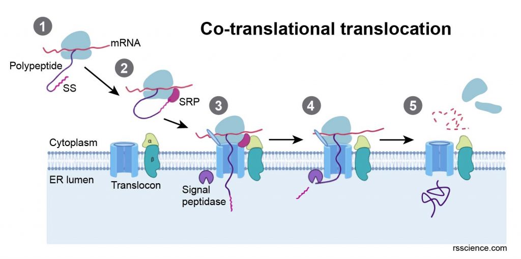
[In this figure] The ribosomes bound to the rough ER’s outer membrane can synthesize new protein directly into the ER lumen. This process is called co-translational translocation: (1) newly translated polypeptide presents a signal sequence (SS), (2) the signal recognition particle (SRP) recognizes SS, (3) the SRP receptor recognizes SRP, allowing the ribosome to bind the translocon and the polypeptide to translocate into the ER lumen, (4) translation resumes and the SS is cleaved by a signal peptidase, (5) translation completes in the ER.
The transportation of proteins from ER to Golgi apparatus (as well as Golgi to lysosomes or cell membrane) is done by the budding of membrane-bound small vesicles from the surface of ER. Part of ER lumen’s content will be enclosed inside the vesicles. These vesicles move in the cytosol by anchoring on the cytoskeleton. Once they arrive at the destination (ie, the cis face of Golgi), the membrane will fuse and the content will be released into the lumen of Golgi cisternae.
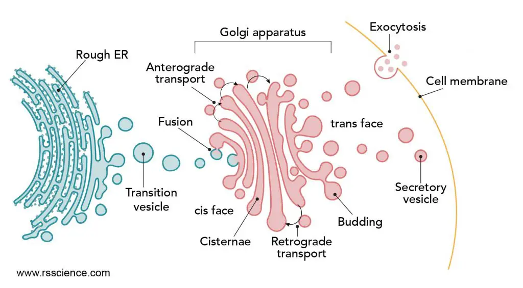
[In this figure] The journey of protein synthesis and transportation.
After proteins are synthesized in the rough ER, they traveled to the Golgi for further glycosylation. Once they are done with the modification, proteins are packed into vesicles, called secretory vesicles. Then, they travel to the membrane and release by exocytosis.
Created with BioRender.com
The proximity of the rough ER to the cell nucleus gives the ER unique control over protein processing. When somethings wrong in protein synthesis and folding occur, misfolded or unfolded proteins accumulate in the ER lumen and thereby influence the overall rate of protein translation. This causes ER stress. A healthy ER can sense the problem and sent a message (known as the unfolded protein response or UPR) to the nucleus. The response is adaptive, such that UPR activation triggers reductions in protein synthesis, enhancements in ER protein-folding capacity, and ER-associated protein degradation. If the rescuing mechanism fails, cells will undergo apoptosis (or programmed cell death).
The function of smooth ER
Smooth ER, by contrast, is not associated with ribosomes, and its functions differ. The smooth ER is involved in the synthesis of lipids, including cholesterol and phospholipids, to produce new cellular membranes. In certain cell types, smooth ER plays an important role in synthesizing steroid hormones from cholesterol. In liver cells, smooth ER contributes to the detoxification of drugs and harmful chemicals.
The sarcoplasmic reticulum (SR) is a specialized type of smooth ER in the cytoplasm of skeletal muscle and heart muscle cells. In these cells, their plasma membrane forms deep invaginations known as T-tubules (or transverse tubules). T-tubules stay close to sarcoplasmic reticulum networks that regulate the calcium ion concentration. This structure allows action potentials to penetrate quickly to the interior of the cell, resulting in maximal muscle force output.
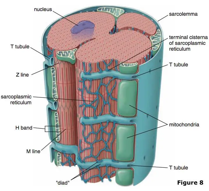
[In this image] T-tubules (transverse tubules) are extensions of the cell membrane that penetrate into the center of skeletal and cardiac muscle cells. T-tubules permit the rapid transmission of the action potential into the cell and also play an important role in regulating cellular calcium concentration.
There are areas where the ER is partly smooth and partly rough, this area is called the transitional ER. The transitional ER gets its name because it contains ER exit sites. The transport vesicles that contain lipids and proteins made in the ER detach from the transitional ER and start moving to the Golgi apparatus.
How big is the endoplasmic reticulum of cells?
The ER is often the largest organelle in eukaryotic cells. If we dissect a eukaryotic cell, the membrane area allocation is dominated by the ER (as much as 60%) followed by the Golgi and mitochondria. Surprisingly, the cell plasma membrane in these mammalian cells tends to be a small fraction of less than 10%.
The ER network can reach the entire cytoplasm. Its cisternae and tubular structures maximize the total membrane surface area for protein and lipid synthesis. This is particularly significant in cell types that are heavily in charge of secreting, like liver and pancreatic cells. In terms of volume, the ER can comprise >10% of the cellular volume.
Endoplasmic reticulum – what does it look like under a microscope?
In the light microscope, the rough endoplasmic reticulum can be seen as masses of material staining with basic dyes. The basophilia of the RER in H&E staining is due to the presence of negatively charged rRNA in the ribosomes attached to the cisternae.
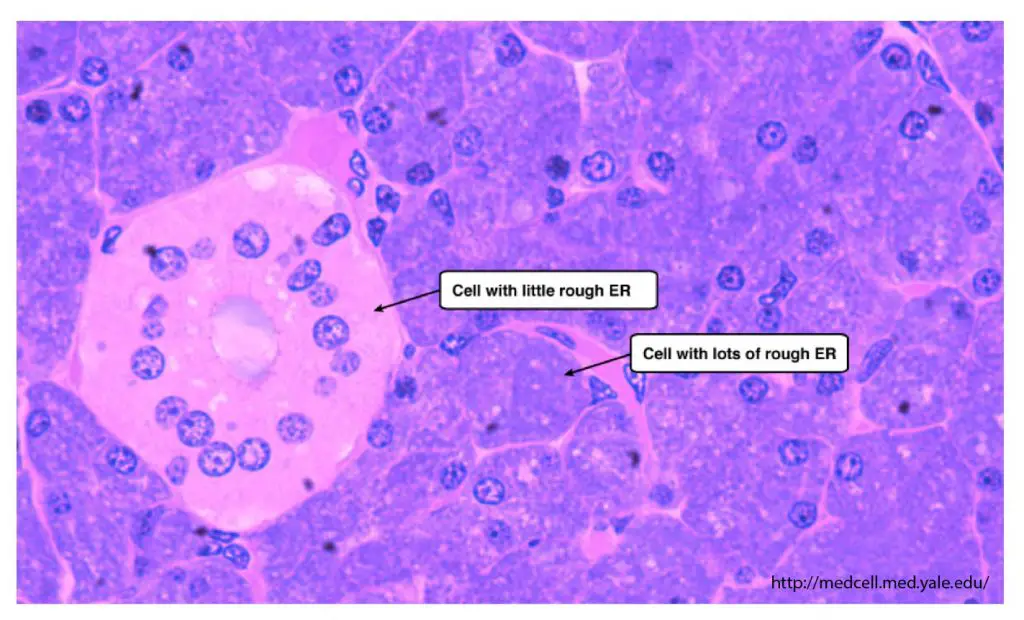
[In this image] Hematoxylin and eosin (H&E) staining of pancreatic tissue.
You can see the cells that produce pancreatic juices (including digestive enzymes) contain abundant rough endoplasmic reticulum. In contrast, cells from the pancreatic ducts don’t need a high amount of RER for protein synthesis.
Photo source: Histology @Yale
Fluorescent microscopy is a powerful tool to study the biology of the endoplasmic reticulum. ER can be labeled by antibodies, lipid dyes, or transgenic GFP proteins.
[In this video] Real-time video of ER (green) and Golgi bodies (magenta) movement in a plant cell is visualized with labeled fluorescent proteins and fluorescent microscopy.
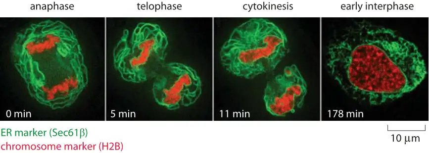
[In this figure] Structural dynamics of the endoplasmic reticulum during the cell cycle.
Photo source: Cell biology by the numbers
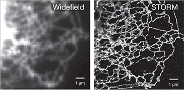
[In this figure] Imaging ER is challenging using the traditional fluorescent microscope (left image, widefield). With new super-resolution microscopy like stochastic optical reconstruction microscopy (STORM), scientists now can study the structure and dynamics in great detail.
Photo source: ThermoFisher Scientific
A transmission electron microscope (TEM) is ideal to see the ultrafine structure of the endoplasmic reticulum.
[In this figure] A transmission electron micrograph (TEM) showing the structure of the endoplasmic reticulum. This is the region surrounding the nucleus in an acinar cell that comes from the pancreas of a bat.
Image source: Cell biology by the numbers
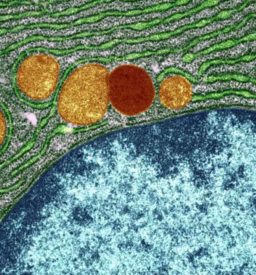
[In this figure] A colored electron micrograph shows the rough endoplasmic reticulum (green) adjacent to the nucleus of a cell (blue).
Credit: University of Edinburgh.
The discovery of ER
The structure of the endoplasmic reticulum was discovered by cell biologists Keith Porter, Albert Claude, and Ernest Fullman, who produced the first electron micrograph of a cell. The name was given by Porter in 1953 to describe the looking of “lace-like reticulum”.
Summary
- Endoplasmic reticulum (ER) is an internal membrane that forms branching networks of many interconnected sacs and tubes.
- There are two types of ER: rough ER and smooth ER.
- The outer side (facing the cytosol) of the rough ER is studded with ribosomes. Under the electron microscope, the dense granular ribosomes gave the name “rough” ER.
- Rough ER stays closer to the nucleus and coordinates protein synthesis.
- Smooth ER lacks ribosomes. It specializes in lipid synthesis, steroid hormone production, and detoxification.
References
“The endoplasmic reticulum: structure, function and response to cellular signaling”
“Endoplasmic Reticulum: Keeping in shape”
“How big is the endoplasmic reticulum of cells?”